
Habit and Habitat
Earthworms are terrestrial annelids found in moist soil, especially rich in organic matter. They prefer loamy and humus-rich soil with sufficient moisture and aeration. It is detritivorous, feeding on decaying organic matter, leaves, and decomposing plant material, helping in soil fertility and decomposition. It is considered a “farmer’s friend” because it helps in vermicomposting, soil aeration, and fertility improvement.
External Features of Pheretima posthuma
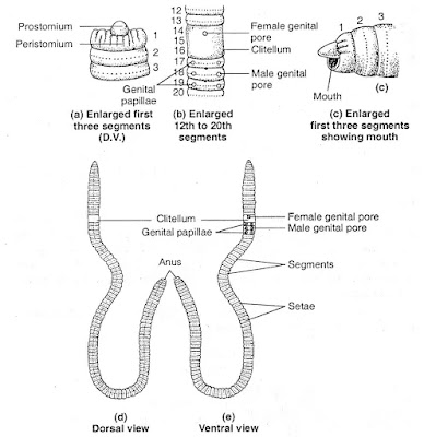
- Shape and Size: Pheretima posthuma has a long, cylindrical, and segmented body. The length ranges from 15 to 30 cm, and the width is about 3–5 mm. The body is bilaterally symmetrical and metamerically segmented (divided into similar ring-like segments).
- Segmentation: The body consists of about 100–120 segments (metameres). Each segment is separated externally by shallow grooves and internally by septa (partitions).
- Color and Body Surface: The body is brownish in color due to the presence of porphyrin pigments, which protect the worm from UV radiation. The dorsal surface has a dark median line due to the presence of a dorsal blood vessel visible beneath the skin. The ventral surface is slightly paler and bears minute bristles called setae that help in locomotion.
- Anterior and Posterior Ends: The anterior end is tapered and pointed, while the posterior end is blunt and rounded. The first segment is called the peristomium, which bears the mouth. The last segment is called the pygidium, which has the anus.
- Clitellum: It is a thick, glandular, saddle-like band present on segments 14–16. It secretes mucus to form a cocoon for fertilized eggs. It is a key feature that differentiates sexually mature worms.
- Setae (Locomotory Structures): Setae are tiny, S-shaped chitinous bristles embedded in the body wall of Pheretima posthuma. They are absent in the first and last segments but are present in all other segments. Each segment (except the peristomium and pygidium) contains about 80–120 setae arranged in rows called parapodia.

- Body Wall: The body wall of Pheretima posthuma has several layers that protect the body and help in movement. The outermost layer is the cuticle, which is thin, transparent, and protects the worm from drying out and infections. Below it is the epidermis, a single layer of cells that produce mucus to keep the body moist for breathing. The muscular layer has two types of muscles: outer circular muscles that help the body stretch and inner longitudinal muscles that help it shorten. These muscles work together to create movement. The coelomic epithelium (parietal peritoneum) is the innermost layer that lines the body cavity, helps in excretion, and protects internal organs.
- Septum: The septum is a thin muscular partition between two adjacent body segments. It is made up of connective tissue and muscle fibers. Septum divides the coelomic cavity into separate chambers in each segment. It provides structural support by maintaining the shape of the worm.
- Coelom (Body Cavity): The coelom is the fluid-filled body cavity present between the gut and body wall. It is a true coelom (schizocoel) lined by coelomic epithelium. It contains coelomic fluid, which is milky white and contains free coelomocytes (immune cells). It acts as a hydrostatic skeleton, providing shape and support.
Digestive System of Earthworm
The digestive system of an earthworm is a well-organized and efficient system adapted for its detritivorous lifestyle. Earthworms feed on decaying organic matter, soil, and microorganisms, and their digestive system is specialized for breaking down this material and extracting nutrients. The digestive system of an earthworm is a straight tube that runs from the mouth to the anus. It consists of the following parts:
- Mouth: The mouth is located on the first segment of the earthworm (peristomium). It is a small opening used for ingesting food, which primarily consists of decaying organic matter, soil, and microorganisms.
- Buccal Cavity: The buccal cavity is a muscular region located just behind the mouth. It helps in the ingestion of food by sucking in soil and organic matter.
- Pharynx: The esophagus is a narrow tube that connects the pharynx to the crop. The pharynx secretes mucus and digestive enzymes, such as amylase, which begin the breakdown of carbohydrates. It transports food from the pharynx to the crop.
- Esophagus: From the pharynx, the food moves into the esophagus, a narrow tube that connects the pharynx to the crop. The esophagus may secrete calcium carbonate to neutralize acidic substances in the soil, protecting the worm’s digestive system.
- Crop: The crop is a thin-walled, sac-like structure located in segments 7 to 9. It serves as a temporary storage organ for food. Food is softened and mixed with digestive enzymes in the crop.
- Gizzard: The gizzard is a thick-walled, muscular organ located in segments 9 to 10. It grinds the food into smaller particles using strong muscular contractions and small soil particles ingested with the food. This grinding action increases the surface area of the food, making it easier for enzymes to act on it.
- Intestine: The intestine is a long, straight tube that extends from the gizzard to the anus. It is the primary site for digestion and absorption of nutrients. The inner lining of the intestine is folded into typhlosole, a dorsal fold that increases the surface area for absorption. The intestine is lined with glandular cells that secrete digestive enzymes, including:
Proteases for protein digestion.
Lipases for fat digestion.
Cellulases for breaking down cellulose (produced by symbiotic microorganisms in the earthworm’s gut).
Nutrients are absorbed through the intestinal wall into the bloodstream for distribution to the rest of the body. - Anus: The anus is located at the posterior end of the earthworm. Undigested material and waste are expelled through the anus in the form of castings (vermicast), which are rich in nutrients and beneficial for soil health.

Excretory System in Earthworm
The excretory system in earthworms is responsible for removing metabolic waste products from the body and maintaining osmotic balance. Earthworms have a specialized excretory system consisting of nephridia, which are tubular structures that function similarly to kidneys in higher animals.
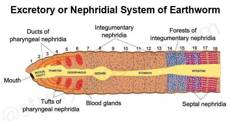
The excretory system in earthworms is composed of nephridia, which are segmentally arranged paired structures. Each segment of the earthworm (except the first three and the last one) contains a pair of nephridia.
Structure of Nephridia
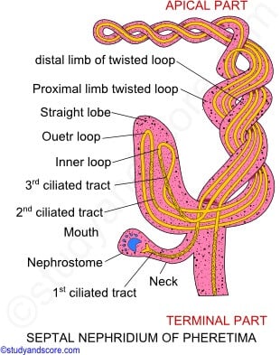
Each nephridium is a tubular structure divided into three main parts:
- Nephrostome: The nephrostome is a funnel-shaped, ciliated opening that collects waste products from the coelomic fluid (fluid in the body cavity). It is located in the anterior part of the nephridium.
- Nephridial Tubule: The nephridial tubule is a long, coiled tube that extends from the nephrostome. It is lined with glandular cells that reabsorb useful substances (e.g., water, ions, and nutrients) from the filtrate.
- Nephridiopore: The nephridiopore is the external opening of the nephridium. It releases the processed waste products (urine) outside the body.
Types of Nephridia
- Septal Nephridia: Septal nephridia are located on the intersegmental septa, which are the walls separating each segment of the earthworm. They are found from the 15th segment to the last segment. Septal nephridia collect waste products from the coelomic fluid and release them into the intestine.The excretory products are then expelled from the body along with digestive waste through the anus. Septal nephridia are the largest and most complex type of nephridia in earthworms.
- Integumentary Nephridia: Integumentary nephridia are located on the inner body wall (integument) of the earthworm. They are found in all segments except the first two segments (peristomium and prostomium). Integumentary nephridia collect waste products from the coelomic fluid and release them directly to the outside through the nephridiopore. Integumentary nephridia are the most numerous type of nephridia in earthworms. They are responsible for the majority of nitrogenous waste excretion.
- Pharyngeal Nephridia: Pharyngeal nephridia are located in the pharyngeal region of the earthworm, near the mouth. They are found in the 4th, 5th, and 6th segments. Pharyngeal nephridia collect waste products from the coelomic fluid and release them into the digestive tract. The excretory products are then expelled from the body along with digestive waste. They are fewer in number compared to septal and integumentary nephridia.
Nervous system of Earthworm
The nervous system of an earthworm is relatively simple but well-organized, allowing it to respond to environmental stimuli and coordinate its movements. It consists of three main components: the central nervous system (CNS), the peripheral nervous system (PNS), and the autonomic nervous system (ANS).

Central Nervous System (CNS)
- Nerve Ring: The nerve ring is a ring-like structure that encircles the pharynx (part of the digestive system) in the third segment of the earthworm. It consists of
a) Supra-pharyngeal Ganglion (Cerebral Ganglion): Located above the pharynx, it acts as the “brain” of the earthworm. It is responsible for receiving sensory input and sending motor signals.
b) Sub-pharyngeal Ganglion: Located below the pharynx, it connects to the supra-pharyngeal ganglion and helps coordinate sensory and motor functions.
The nerve ring integrates sensory information and controls basic behaviors such as feeding and movement. - Ventral Nerve Cord: The ventral nerve cord is a long, double-layered nerve cord that runs along the ventral (bottom) side of the earthworm, from the sub-pharyngeal ganglion to the last segment of the body. It has a ganglion (a cluster of nerve cells) in each segment of the earthworm’s body. These ganglia are connected by longitudinal nerve fibers, which allow for coordinated movement and responses to stimuli. The ventral nerve cord acts as the main pathway for transmitting signals between the brain (supra-pharyngeal ganglion) and the rest of the body.
Peripheral Nervous System (PNS)
The PNS consists of nerves that branch out from the CNS to the rest of the body. It connects the CNS to sensory receptors and muscles.
- a. Sensory Nerves: Sensory nerves carry information from sensory receptors (e.g., photoreceptors, chemoreceptors, and tactile receptors) to the CNS. These receptors help the earthworm detect light, chemicals, touch, and vibrations in its environment.
- b. Motor Nerves: Motor nerves carry signals from the CNS to the muscles, enabling movement and other responses. For example, when an earthworm senses danger, motor nerves trigger muscle contractions to help it burrow into the soil.
Autonomic Nervous System (ANS)
The ANS in earthworms controls involuntary functions, such as digestion and circulation. It works in coordination with the CNS and PNS. It regulates the activity of internal organs, such as the digestive system and circulatory system. For example, it controls the rhythmic contractions of the earthworm’s digestive tract (peristalsis) to move food through the body.
The ANS is connected to the CNS through the ganglia of the ventral nerve cord. It ensures that internal processes are carried out efficiently without conscious control.
Reproductive System of Earthworm
Earthworms are hermaphrodites, meaning each individual possesses both male and female reproductive organs. However, they cannot self-fertilize and require a partner for reproduction. The reproductive system of earthworms is well-organized and involves both male and female structures, as well as specialized processes for cocoon formation and fertilization.

Male Reproductive System
The male reproductive system of earthworms is located in specific segments of their body. It consists of the following structures:
- Testes: Earthworms have two pairs of testes, located in the 10th and 11th segments. These testes produce sperm cells.
- Seminal Vesicles: The sperm produced by the testes mature and are stored in seminal vesicles, which are located in the 11th and 12th segments. These vesicles are large, pouch-like structures that also provide nourishment to the developing sperm.
- Male Genital Pores: The mature sperm are released through male genital pores, which are situated on the 18th segment. During mating, sperm is transferred to the partner through these pores.
Despite having both male and female reproductive organs, earthworms rely on cross-fertilization, where two individuals exchange sperm to fertilize each other’s eggs.
Female Reproductive System
The female reproductive system of earthworms is also located in specific segments and includes the following structures:
- Ovaries: Earthworms have a single pair of ovaries, located in the 13th segment. The ovaries produce eggs.
- Oviducts: The eggs travel from the ovaries through oviducts, which are short tubes that open into the female genital pores.
- Female Genital Pores: The female genital pores are located on the 14th segment. Eggs are released through these pores during reproduction.
- Spermathecae: These are small sac-like structures located in the 6th to 9th segments. They store sperm received from a partner during mating until fertilization occurs.
The presence of both male and female organs in the same individual facilitates efficient reproduction, but cross-fertilization ensures genetic diversity.
Formation of Cocoon
After mating, earthworms form a cocoon to protect the developing embryos. This process involves the following steps:
- Clitellum Secretion: The clitellum, a thickened glandular region located near the anterior end (segments 14th to 16th), secretes a mucous ring.
- Collection of Eggs and Sperm: As the mucous ring slides forward, it collects eggs from the female genital pores and stored sperm from the spermathecae.
- Cocoon Formation: The mucous ring eventually slips off the earthworm’s body and hardens to form a protective cocoon. The cocoon is lemon-shaped and provides a safe environment for the embryos to develop.
The cocoon is a key adaptation that ensures the survival of the offspring by protecting them from environmental hazards and predators.
Fertilization
Fertilization in earthworms occurs externally within the cocoon. The process involves the following steps:
- Sperm Exchange: During mating, two earthworms align themselves in opposite directions and exchange sperm. The received sperm is stored in the spermathecae.
- Egg and Sperm Release: After mating, the eggs and stored sperm are released into the cocoon as the mucous ring slides off the body.
- Fertilization: Fertilization occurs inside the cocoon, where the sperm fertilizes the eggs. The fertilized eggs develop into juvenile earthworms, which eventually emerge from the cocoon.
This method of external fertilization within a protective cocoon ensures the survival and development of the offspring.
Economic Importance of Earthworms
Earthworms are of significant economic and ecological importance due to their role in soil health and agriculture. Their contributions include:
- Soil Fertility: Earthworms enhance soil fertility by breaking down organic matter and converting it into nutrient-rich castings (vermicompost). This process improves soil structure, aeration, and water retention.
- Vermicomposting: Earthworms are widely used in vermicomposting to produce organic fertilizers. This eco-friendly method recycles organic waste and reduces the need for chemical fertilizers.
- Biological Indicators: Earthworms are used as bioindicators to assess soil health and pollution levels. Their presence indicates a healthy, fertile soil ecosystem.
- Fishing Bait: Earthworms are commonly used as bait in fishing due to their availability and attractiveness to fish.