Digestive System in Frog
The digestive system of a frog is well-adapted for its carnivorous diet, primarily consisting of insects, worms, and small aquatic organisms. The system is composed of the alimentary canal and associated digestive glands. The alimentary canal is a continuous tube that runs from the mouth to the cloaca and includes several specialized structures for ingestion, digestion, absorption, and egestion.
Parts of the Alimentary Canal in Frogs:
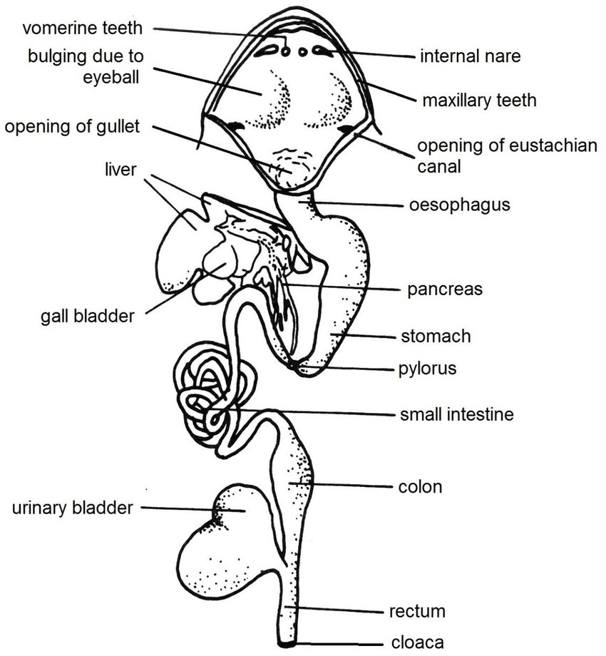
- Mouth: The digestive process begins in the mouth, where the frog captures prey using its sticky tongue. The mouth opens into the buccal cavity, which is lined with mucous membranes and contains small, conical teeth on the upper jaw (maxillary teeth) and a pair of vomerine teeth on the roof of the mouth. These teeth are not used for chewing but for gripping prey.
- Pharynx: The buccal cavity leads to the pharynx, a short, muscular region that connects the mouth to the esophagus. The pharynx plays a role in swallowing and is also involved in respiration.
- Esophagus: The esophagus is a short, narrow tube that transports food from the pharynx to the stomach. It is lined with mucous glands that secrete mucus to lubricate the food, facilitating its passage.
- Stomach: The stomach is a muscular, J-shaped organ that stores and mechanically breaks down food. It has two regions: the cardiac stomach (near the esophagus) and the pyloric stomach (near the small intestine). Gastric glands in the stomach lining secrete digestive enzymes (e.g., pepsin) and hydrochloric acid, which help in the chemical digestion of proteins.
- Small Intestine: The small intestine is the primary site for digestion and absorption of nutrients. It is divided into two parts: the duodenum (upper part) and the ileum (lower part). The duodenum receives bile from the liver and digestive enzymes from the pancreas, which aid in the breakdown of fats, proteins, and carbohydrates. The inner lining of the small intestine has numerous finger-like projections called villi, which increase the surface area for nutrient absorption.
- Large Intestine: The large intestine, also known as the rectum, is a short, wide tube that absorbs water and electrolytes from the undigested food material. It temporarily stores fecal matter before elimination.
- Cloaca: The cloaca is a common chamber that receives undigested waste from the large intestine, as well as urinary and reproductive products. It opens to the exterior through the cloacal aperture, where waste is expelled from the body.
Associated digestive glands
The associated digestive glands in frogs play a vital role in the digestion and absorption of food. These glands secrete enzymes and other substances that break down complex food molecules into simpler, absorbable forms.
- Liver: The liver is the largest gland in the frog’s body and is located just below the diaphragm. The liver produces bile, a greenish-yellow fluid that is stored in the gallbladder. Bile is released into the duodenum (first part of the small intestine) through the bile duct. It helps in the emulsification of fats, breaking them into smaller droplets for easier digestion by lipase. The liver plays a key role in protein, fat, and carbohydrate metabolism.
- Pancreas: The pancreas is a small, irregular, yellowish gland located between the stomach and the duodenum. It is both an exocrine and endocrine gland.
Exocrine Function: The pancreas secretes digestive enzymes into the duodenum through the pancreatic duct.
Endocrine Function: The pancreas contains clusters of cells called Islets of Langerhans, which secrete hormones like insulin and glucagon to regulate blood sugar levels. - Gastric Glands: These glands are located in the lining of the stomach. They secrete gastric juice, which contains:
- Hydrochloric Acid (HCl): Creates an acidic environment for enzyme activity and kills bacteria in food. Pepsinogen: An inactive enzyme that is converted to pepsin in the presence of HCl. Pepsin breaks down proteins into smaller peptides.
- Intestinal Glands: These glands are found in the lining of the small intestine. They secrete intestinal juice (succus entericus), which contains enzymes like maltase, sucrase, and lipase. The intestinal glands also secrete mucus to protect the intestinal lining and facilitate the movement of food.
- Mucous Glands: These glands are present in the buccal cavity, esophagus, and stomach. They secrete mucus, which lubricates food and protects the lining of the digestive tract from mechanical damage and digestive enzymes.
Physiology of Digestion in Frog
Frogs are carnivorous as adults, feeding primarily on insects, spiders, earthworms, snails, fish, small frogs, and other prey. Their digestive system is adapted to efficiently process these types of food.
Ingestion and Mechanical Digestion
- Mouth and Buccal Cavity: Frogs use their sticky tongue to capture prey. The tongue is attached at the front of the mouth and flips out rapidly to catch insects or other small animals. The maxillary teeth (on the upper jaw) and vomerine teeth (on the roof of the mouth) help grip the prey but do not chew it. Frogs swallow their prey whole. Salivary glands secrete mucus to lubricate the food, making it easier to swallow.
- Esophagus: The food passes from the mouth to the stomach via the esophagus. The esophagus is short and wide, allowing for rapid passage of food.
Chemical Digestion:
- Stomach: The stomach is a muscular organ that stores and digests food. Gastric glands in the stomach secrete hydrochloric acid (HCl) and the enzyme pepsinogen (which converts to pepsin in the acidic environment). Pepsin breaks down proteins into smaller peptides. The churning action of the stomach (mechanical digestion) mixes the food with digestive juices, forming a semi-liquid mixture called chyme.
- Small Intestine: The chyme moves from the stomach to the small intestine, where most of the digestion and absorption occur. The liver secretes bile, which is stored in the gallbladder and released into the small intestine. Bile emulsifies fats, breaking them into smaller droplets for easier digestion. The pancreas secretes digestive enzymes such as:
a) Trypsin and chymotrypsin: Break down proteins into amino acids.
b) Amylase: Breaks down carbohydrates into simple sugars.
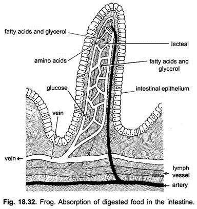
c) Lipase: Breaks down fats into fatty acids and glycerol.
Absorption:
The inner lining of the small intestine is folded into villi and microvilli, which increase the surface area for absorption. Nutrients such as amino acids, simple sugars, and fatty acids are absorbed into the bloodstream through the intestinal walls.
Large Intestine and Egestion:
- Large Intestine: The remaining undigested material passes into the large intestine, where water and electrolytes are reabsorbed. The waste material from digestion is compacted into fecal pellets.
- Cloaca: The large intestine opens into the cloaca, a common chamber for the digestive, excretory, and reproductive systems. Waste material is expelled through the cloacal opening in the form of feces.
Circulatory system in Frog
Frogs have a closed circulatory system with a three-chambered heart and a double circulation pattern. Their circulatory system efficiently transports oxygen, nutrients, and waste throughout the body.
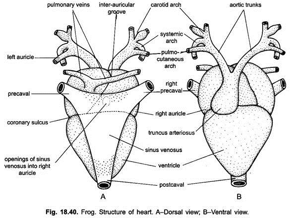
Heart: The frog’s heart has three chambers: two atria and one ventricle. The right atrium receives deoxygenated blood from the body, while the left atrium receives oxygenated blood from the lungs and skin. The ventricle pumps mixed blood to different parts of the body.
- External Features: The heart is triangular in shape and reddish in color. It is enclosed in a double-walled pericardium filled with pericardial fluid, which protects and lubricates the heart. Major blood vessels (arteries and veins) are connected to the heart for circulation.
- Internal Structure: The frog’s heart is composed of three chambers.
a) Two Atria (Auricles): They are upper chambers of the heart. The right atrium receives deoxygenated blood from the body through the sinus venosus and vena cava, while the left atrium receives oxygenated blood from the lungs and skin via the pulmonary veins. These two atria are separated by an interatrial septum and function independently to collect and transfer blood into the single ventricle.
b) One Ventricle: The ventricle, which has thick muscular walls, pumps blood to different parts of the body through the conus arteriosus. Although there is no complete separation of oxygenated and deoxygenated blood in the ventricle, muscular folds help direct the flow, minimizing mixing. This structure allows frogs to maintain an efficient double circulation system, ensuring proper oxygenation despite having a three-chambered heart.
Associated Structures:
- Sinus Venosus: A thin-walled, triangular sac located behind the heart. It collects deoxygenated blood from major veins (anterior and posterior vena cava) and directs it into the right atrium.
- Conus Arteriosus (Truncus Arteriosus): A narrow, muscular tube extending from the ventricle. It contains a spiral valve, which helps in directing blood flow to different arteries.
Arterial system in frog
The arterial system in frogs is responsible for distributing oxygenated blood from the heart to various body parts. It originates from the truncus arteriosus, which arises from the ventricle of the heart and divides into three pairs of major arterial branches. These arteries ensure proper circulation and supply oxygenated blood to the head, limbs, and visceral organs.
- Truncus Arteriosus: The arterial system in frogs begins with the truncus arteriosus, a short, thick-walled vessel that arises from the ventricle of the heart. It divides into two branches, each further splitting into three major arches: carotid arch, systemic arch, and pulmocutaneous arch.
- Carotid Arch (Common Carotid Artery): This arch supplies blood to the head, brain, eyes, and tongue.It divides into two major arteries:
External Carotid Artery: Supplies blood to the tongue and buccal cavity.
Internal Carotid Artery: Carries blood to the brain.
The carotid labyrinth, a small dilated structure, helps regulate blood pressure.
- Systemic Arch (Aortic Arch): These arches are the largest and most important, supplying blood to most of the body. The right and left systemic arches curve backward and unite dorsally to form the dorsal aorta. Major branches from the systemic arch include:
Subclavian Artery: Supplies blood to the forelimbs.
Celiacomesenteric Artery: Provides blood to the stomach, intestines, pancreas, and liver.
Renal Arteries: Supply blood to the kidneys for filtration.
Dorsal Aorta (continuation of systemic arches): Further divides into iliac arteries to supply the hind limbs.
- Pulmocutaneous Arch: This arch carries deoxygenated blood to the lungs and skin for gas exchange. It divides into:
Pulmonary Artery: Transports blood to the lungs for oxygenation.
Cutaneous Artery: Supplies blood to the skin, facilitating cutaneous respiration, especially when the frog is submerged in water.

Source: https://commons.wikimedia.org/wiki/File:Artery_system_in_frog.jpg
Venous System of Frog
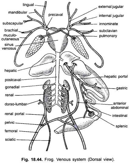
The venous system in frogs consists of a network of veins that collect deoxygenated blood from various parts of the body and return it to the heart. It consists of three major sets of veins: pulmonary veins, systemic veins, and portal veins.
- Pulmonary Veins: These veins carry oxygenated blood from the lungs to the left atrium of the heart. There are two pulmonary veins, one from each lung, which unite before entering the heart. This vein plays a crucial role in pulmonary circulation, ensuring oxygenated blood reaches the systemic circulation.
- Systemic Veins: Systemic veins collect deoxygenated blood from different parts of the body and transport it back to the heart. The main systemic veins include:
Precaval Veins (Anterior Vena Cava): Two large veins that collect blood from the head, forelimbs, and upper body. Each precaval vein is formed by the union of:
External Jugular Vein: It receives blood from the tongue (lingual) and floor of mouth (mandibular).
Innominate Vein: It receives branches from the brain and orbit (internal jugular), and fromshoulder and back of arm (subscapular).
Subclavian Vein: It receives branches from arm (brachial) and from skin and muscles of the abdomen and also mucous membrane of mouth and head muscles (musculo-cutaneous)
Postcaval Vein: It is a large vein that drains deoxygenated blood from the posterior part of the body and empties into the sinus venosus. It is formed by the union of several major veins, including:
Renal Veins: It collects blood from the kidneys after filtration.
Gonadal Veins: These veins are responsible for draining blood from the testes in males (spermatic veins) and ovaries in females (ovarian veins).
Hepatic Veins: It collects blood from the liver. - Portal Veins: Portal veins play a crucial role in filtering blood before it reaches the heart. The two major portal systems in frogs are:
Hepatic Portal System: Blood from the stomach, intestines, pancreas, and spleen is first collected by the hepatic portal vein, which then passes through the liver for detoxification before draining into the postcaval vein.
Renal Portal System: Blood from the hind limbs and lower body is collected by the renal portal veins, which pass through the kidneys before entering the postcaval vein. This helps in the excretion of nitrogenous wastes.
Reproductive system of frog
The reproductive system in frogs is well-adapted for external fertilization and is distinct between males and females.
Male Reproductive System
The male reproductive system in frogs is responsible for producing and delivering sperm. It consists of the following parts:
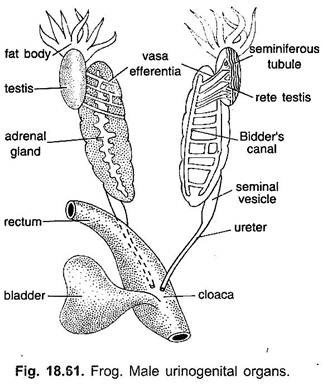
- Testes: The testes are paired, oval-shaped organs located near the kidneys. They produce sperm (male gametes) and the hormone testosterone, which regulates male reproductive behavior and secondary sexual characteristics.
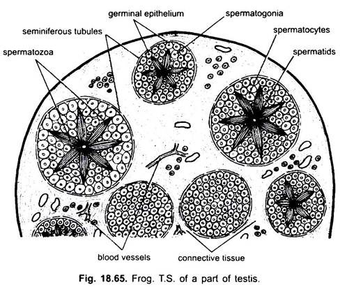
The transverse section (T.S.) of the testes in a frog shows its internal structure and sperm-producing components. The testes are made up of numerous seminiferous tubules, which are lined with germinal epithelium. These tubules produce spermatogonia, which undergo development into mature spermatozoa through different stages like spermatocytes and spermatids. The spaces between the tubules contain Leydig cells, which produce hormones that regulate reproduction. The mature sperm move into the vasa efferentia, which transport them to the Bidders’ canal and then to the ureter for release during reproduction.
- Vasa Efferentia: The vasa efferentia are thin tubes that originate from the inner edge of the testis and extend through the mesorchium before reaching the kidney. There, they connect to the Bidder’s canal, which is linked to the ureter through the kidney’s collecting tubules. This pathway allows sperm to travel from the vasa efferentia into the Bidder’s canal, then through the collecting tubules, and finally into the ureter.
- Kidneys: The kidneys are part of the excretory system but also play a role in reproduction. In male frogs, the kidneys help transport sperm from the vasa efferentia to the ureters.
- Ureters (Urinogenital Ducts): A common chamber at the end of the digestive, excretory, and reproductive tracts. It receives sperm from the ureters and releases it during mating.
- Copulatory Pad (Nuptial Pad): It is present on the inner side of the forelimbs in some male frogs. It helps the male grasp the female during amplexus (mating).
Female Reproductive System
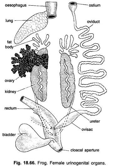
The female reproductive system of a frog consists of paired ovaries, oviducts, and cloaca, which work together to produce and transport eggs.
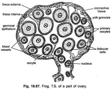
- Ovaries: A female frog has two ovaries, located near the kidneys. They contain numerous eggs at different stages of development. During the breeding season, the ovaries enlarge and fill with mature eggs.
The transverse section (T.S.) of the ovary in a frog shows its structure and different stages of egg development. The outer germinal epithelium produces egg cells, while connective tissue and blood vessels provide nutrients. Inside, numerous oocytes (developing eggs) are present at various stages , smaller ones near the wall and larger, yolk-filled mature eggs towards the center. Each egg is surrounded by follicular cells that aid in nutrient supply. The central ovarian lumen contains fluid and helps in ovulation. During the breeding season, the ovary enlarges, and mature eggs are released into the oviduct for fertilization.
- Oviducts: Each ovary is connected to a long, coiled oviduct. The funnel-shaped opening (ostium) at the front end of the oviduct collects the released eggs. As eggs pass through the oviduct, they get coated with a jelly-like substance, which protects them and helps in external fertilization.
- Uterus (Ovisac): The lower part of the oviduct expands into a temporary storage chamber for eggs before they are laid.
- Cloaca: The oviducts open into the cloaca, a common chamber for the reproductive, excretory, and digestive systems. The oviducts open into the cloaca, a common chamber for the reproductive, excretory, and digestive systems. During spawning, eggs are released from the cloaca into water, where external fertilization takes place with male sperm.